Dog Leg Bone Diagram : A Visual Guide To Dog Anatomy Muscle Organ Skeletal Drawings All Things Dogs All Things Dogs / The foot bones shown in this diagram are the talus, navicular, cuneiform, cuboid, metatarsals and calcaneus.
Dog Leg Bone Diagram : A Visual Guide To Dog Anatomy Muscle Organ Skeletal Drawings All Things Dogs All Things Dogs / The foot bones shown in this diagram are the talus, navicular, cuneiform, cuboid, metatarsals and calcaneus.. The bones and muscles of the rear leg showing the joints. Carpal joint the carpal (wrist) joint is where the radius and ulna join with seven small carpal bones. In addition to its structural functions (keeping the dog from falling and facilitating locomotion), this system of joint and bones is capable of performing both generalized and highly specific movements. Hock joint the hock (ankle) joint connects the paw (talus and calcaneus bones) to the shin bones (tibia and fibula). What are large leg bones?
The foot bones shown in this diagram are the talus, navicular, cuneiform, cuboid, metatarsals and calcaneus. What is the anatomy of a dog's leg? Bone of the paw of the foreleg between the carpus and the phalanges. These further extend to the heel bone, known as tarsus, the paw bone, known as metatarsus, and the toe bone, phalange. They have small, tight feet, walking on their toes;
Canine Anatomy Veterian Key from i1.wp.com A joint is formed when two bones are brought together and held in place by supporting tissue. These further extend to the heel bone, known as tarsus, the paw bone, known as metatarsus, and the toe bone, phalange. Pain, swelling and heat associated with the affected joint are indications of the condition. Uppermost part of the rear leg of a dog. Hyaluronic acid provides lubrication to the synovial membrane surface and together with another protein, lubricin, it also lubricates the articular cartilage. The elbow is located below the chest at the back of the foreleg. Bone of the paw of the foreleg between the carpus and the phalanges. In synovial joints, resilience of cartilage tissue is important for normal motion as well as shock absorption.
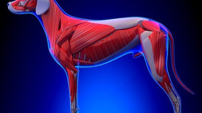
The bones and muscles of the rear leg showing the joints.
It is made of the ulna and the radius. The bones and muscles of the rear leg showing the joints. The bone between the hip and knee is the femur. What is the anatomy of a dog's leg? From the carpal bones ensue five metacarpal bones which connect to the bones of the foot, termed the phalanges. Jul 15, 2021 · dog leg bone diagram / dog anatomy leg bones stock image stock photo download image now istock / paw bone between the heel and the phalanges. Nov 12, 2018 · dog hind leg anatomy. The hind leg can be confusing to some owners, but it has some of the same features as a human. Dog leg anatomy is complex, especially dog knees, which are found on the hind legs. Joints may have ranges of movement such as the shoulder and hip joints, or have very little movement such as joints between the bones in the skull. Uppermost part of the rear leg of a dog. Hock instability results in a sudden onset of lameness. What are the body parts of a dog?
The foot bones shown in this diagram are the talus, navicular, cuneiform, cuboid, metatarsals and calcaneus. What are the body parts of a dog? The forearm is the long bone that runs just after the elbow. The bone between the hip and knee is the femur. They have small, tight feet, walking on their toes;
Anatomy Of Dog Paws With Forelimb And Hindlimb Bones Vector Illustration Dog Anatomy Anatomy Dog Paws from i.pinimg.com The hock is like the human ankle. Ankylosis (stiffness of a joint due to abnormal adhesion and rigidity of the bones of the joint) of the carpal joints can be exceptionally negative for the dog, seriously limiting standing capacities and lateral locomotion. The elbow is located below the chest at the back of the foreleg. Carpal joint the carpal (wrist) joint is where the radius and ulna join with seven small carpal bones. It is made of the ulna and the radius. The bone between the hip and knee is the femur. This short post will try to cover the dog leg anatomy in detail with labeled diagrams. One of two bones of the lower rear leg of a dog.
Below the knee is the tibia and fibula.
Dogs have disconnected shoulder bones (lacking the collar bone of the human skeleton) that allow a greater stride length for running and leaping. The hock is like the human ankle. What are large leg bones? Below the knee is the tibia and fibula. Bone of the paw of the foreleg between the carpus and the phalanges. Nov 12, 2018 · dog hind leg anatomy. Carpal joint the carpal (wrist) joint is where the radius and ulna join with seven small carpal bones. They have small, tight feet, walking on their toes; Hock instability results in a sudden onset of lameness. The technical term for a dog knee is the stifle joint. You've probably heard this mentioned more in horses. What causes cancer in dogs legs? From the carpal bones ensue five metacarpal bones which connect to the bones of the foot, termed the phalanges.
What causes cancer in dogs legs? The bone between the hip and knee is the femur. One of two bones of the lower rear leg of a dog. Hyaluronic acid provides lubrication to the synovial membrane surface and together with another protein, lubricin, it also lubricates the articular cartilage. This is the first joint in the leg.
A Visual Guide To Dog Anatomy Muscle Organ Skeletal Drawings All Things Dogs All Things Dogs from www.allthingsdogs.com Bone of the paw of the foreleg between the carpus and the phalanges. They have small, tight feet, walking on their toes; Then we get to the hock; The bones and muscles of the rear leg showing the joints. This is the first joint in the leg. Dog leg anatomy is complex, especially dog knees, which are found on the hind legs. What are the body parts of a dog? The forearm is the long bone that runs just after the elbow.
Dog leg anatomy is complex, especially dog knees, which are found on the hind legs.
The technical term for a dog knee is the stifle joint. Then we get to the hock; Paw bone between the heel and the phalanges. Jul 09, 2021 · anatomically, the term leg means the part of the hind limb that extends from the stiffle joint to the hock joint (knee to ankle or tibia and fibula bones region). The rear legs of the dog begin with the femur bone, which extends to a pair of bones known as the tibia and the fibula. Their rear legs are fairly rigid and sturdy; Hock instability can occur due to tearing of ligaments that hold the bones of the hock in place, or bone fractures. What are the body parts of a dog? What causes cancer in dogs legs? Pain, swelling and heat associated with the affected joint are indications of the condition. Dogs have disconnected shoulder bones (lacking the collar bone of the human skeleton) that allow a greater stride length for running and leaping. The bone between the hip and knee is the femur. Hyaluronic acid provides lubrication to the synovial membrane surface and together with another protein, lubricin, it also lubricates the articular cartilage.
Hock instability results in a sudden onset of lameness leg bone diagram. License image the bones of the leg are the femur, tibia, fibula and the foot bones shown in this diagram are the talus, navicular, cuneiform, cuboid, metatarsals and from dogs with three legs to cats without eyes, the perfect imperfection photo series celebrates the.


0 Komentar